|
Hybrid CT™ - THE NEW HYBRID COMPUTER TOMOGRAPH FAMILY
 In winning the 2007 Innovation Prize for the NanoSPECT/CT high-resolution imaging system – an instrument used worldwide in pharmaceutical experiments on small animals – MEDISO proved that it is well on the way towards developing hybrid computer tomographs. In winning the 2007 Innovation Prize for the NanoSPECT/CT high-resolution imaging system – an instrument used worldwide in pharmaceutical experiments on small animals – MEDISO proved that it is well on the way towards developing hybrid computer tomographs.
Building upon this preclinical success, doctors at the Institute of Nuclear Medicine at the Medical Faculty of Pecs University have been successfully using the first human SPECT/CT camera produced by MEDISO for more than half of a year.
The non-invasive, hybrid 3D imaging techniques employed by these systems represent the future of diagnostic procedures.
The well-known method of fusing separately-acquired SPECT and CT images is a proven diagnostic tool that has been in use for several years. The combination of SPECT and CT constitutes a versatile and economic imaging procedure, simultaneously showing function and anatomical information of organs and other structures.
Further, an examination conducted with hybrid equipment is more comfortable for a patient as only one short, painless acquisition session is required to obtain both SPECT and CT data. It is also beneficial for the physician as the fused images from the two modalities – collected with the patient in exactly the same position – enable a more precise diagnosis, eliminating the confusion and artifacts that often arise when trying to combine independently-collected images.
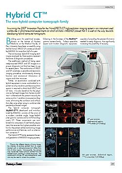
The hybrid computer tomograph (Hybrid CT™) developed and manufactured by MEDISO, consists of the union of a modern, variable angle, large field-of-view gamma camera and a multislice diagnostic CT. These components are combined into one piece of user-friendly equipment that ensures the highest possible comfort for the patient while maintaining image performance and features, such as attenuation correction.*
The Hybrid CT™ family represents the second generation of MEDISO imaging systems, following in the footsteps of the Nucline™ gamma camera family. Today’s specialists expect such modern diagnostic equipment, capable of providing the precise information needed for early diagnosis, thus significantly increasing the possibility of recovery.
*Due to the different density of living tissues, the intensity of photons emitted from an organ labeled with a radiopharmaceutical differs from that of other organs. Consequently, the camera may imprecisely detect photons emitted from the labeled organ, resulting in a false diagnosis. The combination of X-ray CT and mathematical methods may be used to correct this problem.


| 


 Súgó
Súgó